Recent


Researchers map gene activity in excitatory neurons and microglia, offering new insight into depression's biological basis and pointing to future treatments.

October 11, 2025
Source:
ScienceDaily
Breakthrough in Depression Research
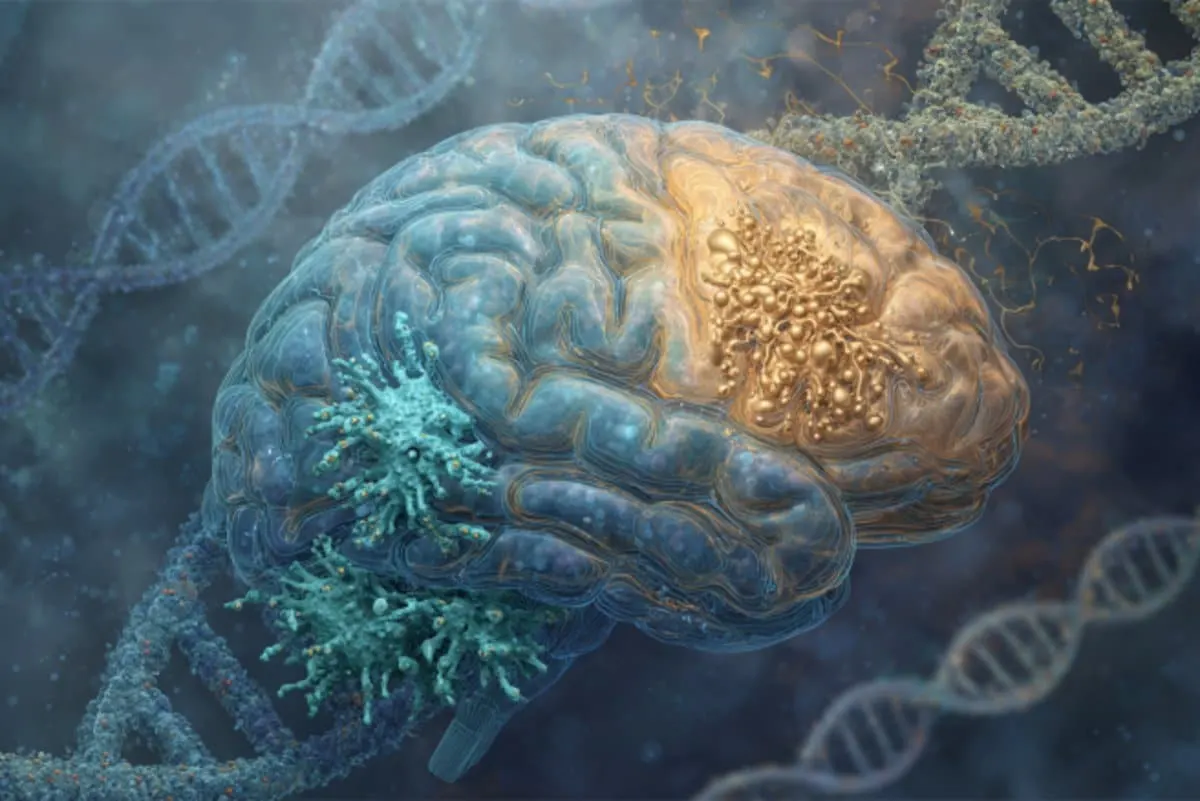
A study published in Nature Genetics has pinpointed specific brain cells that are significantly altered in people suffering from depression. Scientists from McGill University and the Douglas Research Centre led this collaborative effort, employing state-of-the-art single-cell genomics on post-mortem brain samples from 100 individuals. The project's scale and technology represent a milestone in mental health research (McGill News).
Key Findings
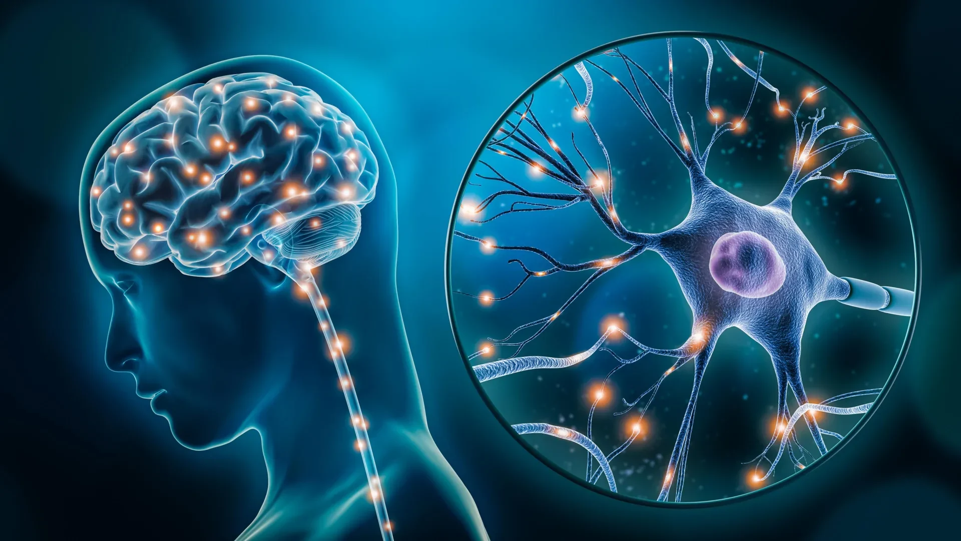
Altered gene expression was found in both excitatory neurons, involved in mood and stress, and microglia, the brain's immune cells.
Evidence of biological change demonstrates depression has a measurable cellular basis, moving understanding beyond psychological models.
Potential for new therapies as researchers now know which cell types and pathways are most affected.
Keep up with the story. Subscribe to the PR+ free daily newsletter
Source:
Neuroscience News
How Brain Cell Alterations Impact Depression
Excitatory Neurons and Microglia
Excitatory neurons regulate mood and response to stress. Disrupted gene activity here may contribute directly to depressive symptoms (NCBI).
Microglia, as immune cells, manage inflammation in the brain. Their abnormal activation can trigger neuroinflammation, damaging neurons and disrupting synaptic connections.
Cycle of Neuronal Damage
Abnormal microglial activation may worsen depression by creating a feedback loop of neuronal damage and chronic inflammation, a phenomenon discussed in recent psychiatric research (Frontiers in Cellular Neuroscience).
Read More


Source:
Journal of Neuroinflammation - BioMed Central
Future Directions and Wider Implications
Therapeutic and Diagnostic Advances
Identifying exact molecular and cell-type changes opens doors to more targeted treatments for depression.
The findings may also help in developing biomarkers for use in living patients, moving toward more personalized medicine (Douglas Research Centre).
Broader Impact
This discovery calls attention to the role of neuroinflammation and immune signaling in psychiatric disorders, a growing focus in mental health science.
Researchers stress the complexity of depression, noting that both genetic and environmental factors influence these cellular pathways.
“This is the first time we’ve been able to identify what specific brain cell types are affected in depression by mapping gene activity together with mechanisms that regulate the DNA code.” — Dr. Gustavo Turecki, senior author
How do the altered gene activities in neurons and microglia specifically contribute to depression?
Altered genes in excitatory neurons affect mood and stress regulation, while changes in microglia can cause neuroinflammation, which disrupts neurons and can lead to depressive symptoms.
What are the potential therapeutic targets identified in this study?
How does the activation of microglia lead to neuroinflammation in depressed individuals?
What role do excitatory neurons play in mood regulation and stress management?
How does the genomic mapping of post-mortem brain tissue differ from other research methods?
Share this news: